Summary information and primary citation
- PDB-id
-
1tup;
DSSR-derived features in text and
JSON formats
- Class
- antitumor protein-DNA
- Method
- X-ray (2.2 Å)
- Summary
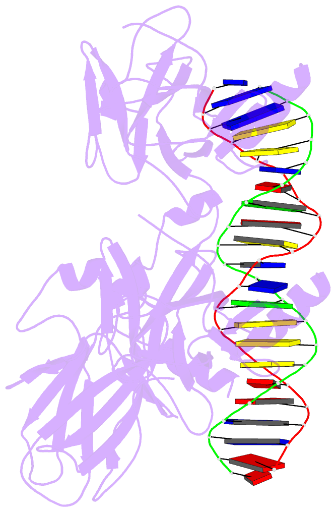
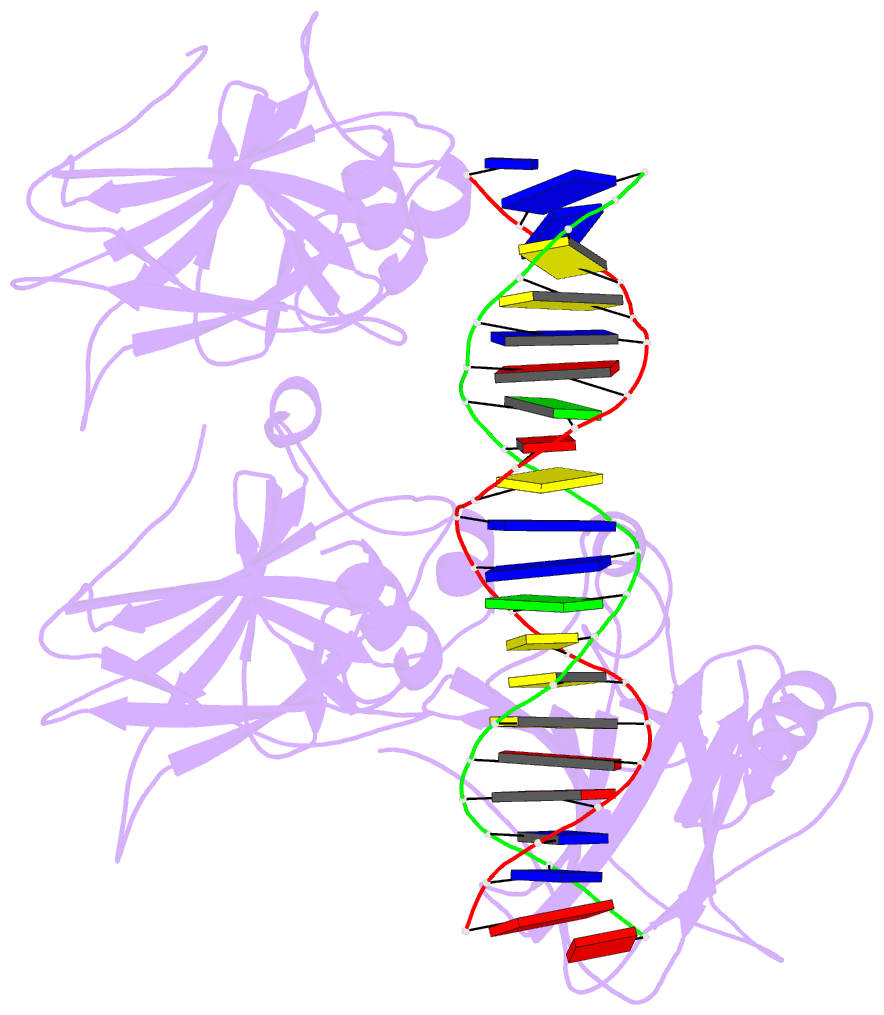
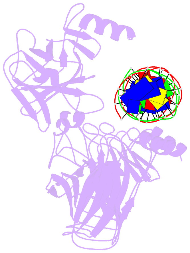
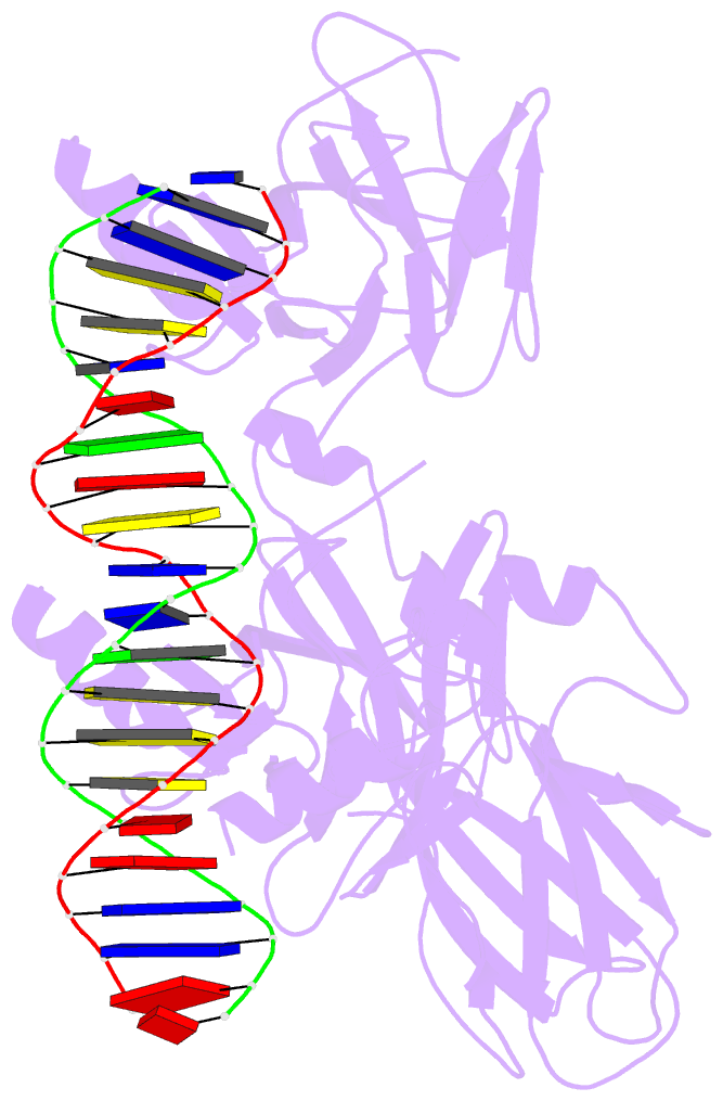
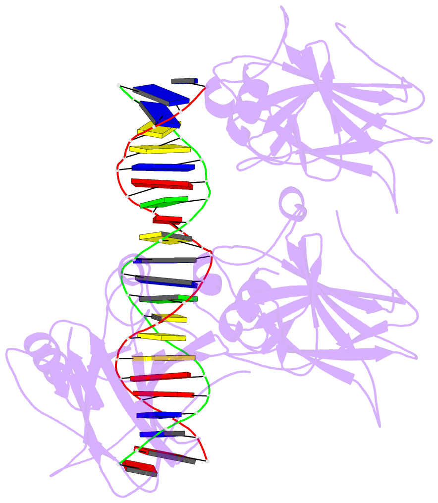
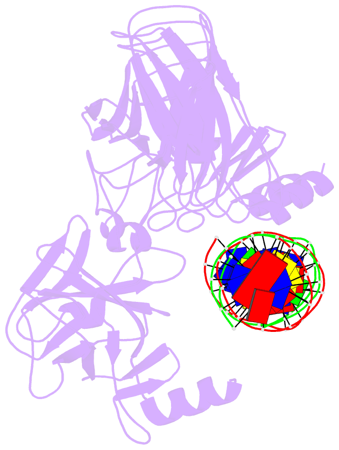
- Tumor suppressor p53 complexed with DNA
- Reference
-
Cho Y, Gorina S, Jeffrey PD, Pavletich NP (1994):
"Crystal
structure of a p53 tumor suppressor-DNA complex:
understanding tumorigenic mutations."
Science, 265, 346-355.
- Abstract
- Mutations in the p53 tumor suppressor are the most
frequently observed genetic alterations in human cancer.
The majority of the mutations occur in the core domain
which contains the sequence-specific DNA binding activity
of the p53 protein (residues 102-292), and they result in
loss of DNA binding. The crystal structure of a complex
containing the core domain of human p53 and a DNA binding
site has been determined at 2.2 angstroms resolution and
refined to a crystallographic R factor of 20.5 percent. The
core domain structure consists of a beta sandwich that
serves as a scaffold for two large loops and a
loop-sheet-helix motif. The two loops, which are held
together in part by a tetrahedrally coordinated zinc atom,
and the loop-sheet-helix motif form the DNA binding surface
of p53. Residues from the loop-sheet-helix motif interact
in the major groove of the DNA, while an arginine from one
of the two large loops interacts in the minor groove. The
loops and the loop-sheet-helix motif consist of the
conserved regions of the core domain and contain the
majority of the p53 mutations identified in tumors. The
structure supports the hypothesis that DNA binding is
critical for the biological activity of p53, and provides a
framework for understanding how mutations inactivate
it.