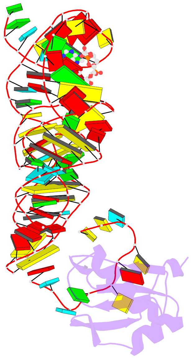
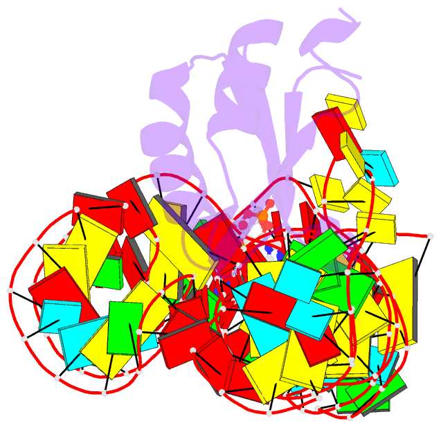
Summary information and primary citation
- PDB-id
-
3muv;
DSSR-derived features in text and
JSON formats
- Class
- RNA binding protein-RNA
- Method
- X-ray (3.2 Å)
- Summary
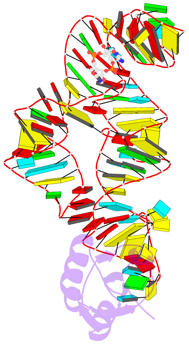
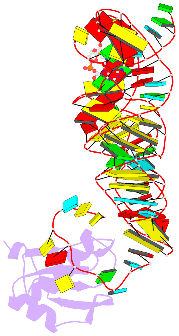
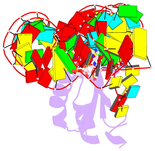
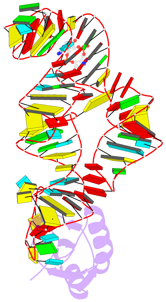
- Crystal structure of the g20a-c92u mutant c-di-gmp
riboswith bound to c-di-amp
- Reference
-
Smith KD, Lipchock SV, Livingston AL, Shanahan CA,
Strobel SA (2010): "Structural
and biochemical determinants of ligand binding by the
c-di-GMP riboswitch ." Biochemistry,
49, 7351-7359. doi: 10.1021/bi100671e.
- Abstract
- The bacterial second messenger c-di-GMP is used in many
species to control essential processes that allow the
organism to adapt to its environment. The c-di-GMP
riboswitch (GEMM) is an important downstream target in this
signaling pathway and alters gene expression in response to
changing concentrations of c-di-GMP. The riboswitch
selectively recognizes its second messenger ligand
primarily through contacts with two critical nucleotides.
However, these two nucleotides are not the most highly
conserved residues within the riboswitch sequence. Instead,
nucleotides that stack with c-di-GMP and that form tertiary
RNA contacts are the most invariant. Biochemical and
structural evidence reveals that the most common natural
variants are able to make alternative pairing interactions
with both guanine bases of the ligand. Additionally, a
high-resolution (2.3 A) crystal structure of the native
complex reveals that a single metal coordinates the
c-di-GMP backbone. Evidence is also provided that after
transcription of the first nucleotide on the 3'-side of the
P1 helix, which is predicted to be the molecular switch,
the aptamer is functional for ligand binding. Although
large energetic effects occur when several residues in the
RNA are altered, mutations at the most conserved positions,
rather than at positions that base pair with c-di-GMP, have
the most detrimental effects on binding. Many mutants
retain sufficient c-di-GMP affinity for the RNA to remain
biologically relevant, which suggests that this motif is
quite resilient to mutation.